Kathryn from New Jersey asked me if she could wait to have a root canal treatment. She had a tooth that was recently filled, then went to a new dentist who said that there was something on the x-ray that looked like decay under the filling, therefore she needed a root canal treatment. See the earlier post: “There are a lot of things that can look like decay on an x-ray.”
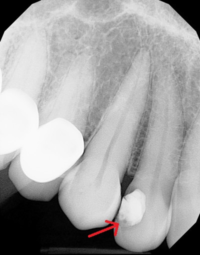
Here is the x-ray she sent, with the tooth in question marked:

And here are my comments:
Kathryn,
Maybe there needs to be more training in dental school on radiographic diagnosis. This is by no means a case calling for plunging in and doing a root canal treatment.
Yes, I see the dark area under the filling, but I see three characteristics of this dark area that they are not telling you about:
1) While tooth decay will show up as a dark area, not all dark areas are decay. This could be a radiolucent base material under the filling, a gap in the filling, or decay – three different possibilities.
2) The dark area is on the surface of the tooth. If it is decay, the dentist should be able to poke that area with an explorer and it will be soft. You gave me no indication that they did this because you said, “There is what looks like some decay under the filling on x-rays.”
3) The dark area does not go anywhere near the pulp. Now there is an optical illusion that creates the impression that the dark area is directly under the filling and between the filling and the pulp. The way to deal with that is to cover up the filling, which is white, with your finger. Now look at the remaining tooth structure. When you do that, you can see the clearly defined margins of the dark area (decay would have fuzzy edges) and that they stop well short of the pulp. There are a good 2 to 3 millimeters of solid tooth structure between the filling and the pulp, with no radiolucent area except that which is on the surface of the tooth, which is also a good distance from the pulp.
Now I can’t give a definitive diagnosis here without actually seeing your tooth and finding out more about what went on, but I can tell you that if they are just relying on the x-ray, there is no justification for doing a root canal treatment. There is no indication of inflammation near the root tip, which is the first indication of an infected tooth that can be seen on an x-ray. And you are saying that the tooth feels fine.
Finally, there is some clear evidence on the x-ray that the root of the tooth is healthy.
If you look at a clearly healthy tooth – for example, the canine tooth next to this one. There is a thin white line that goes all the way around the root of the tooth. This is called the lamina dura. When a tooth gets infected, one of two things happens to this white line. It either becomes broken in the area around the root tip, or it pulls away from the tooth. When I look at the lamina dura of your lateral incisor, I can follow it all the way around the root tip. Because of the angle, the lamina dura appears more fuzzy than it does on the canine, but it is nonetheless there and intact. THAT is the key to the diagnosis of this tooth. The root is healthy.
Dr. Hall
| We thank our advertisers who help fund this site. |
About David A. Hall
Dr. David A. Hall was one of the first 40 accredited cosmetic dentists in the world. He practiced cosmetic dentistry in Iowa, and in 1990 earned his accreditation with the American Academy of Cosmetic Dentistry. He is now president of Infinity Dental Web, a company in Mesa, Arizona that does advanced internet marketing for dentists.
